
Hippocampus-focused VGG16 for CN / MCI / AD classification from T1 MRI
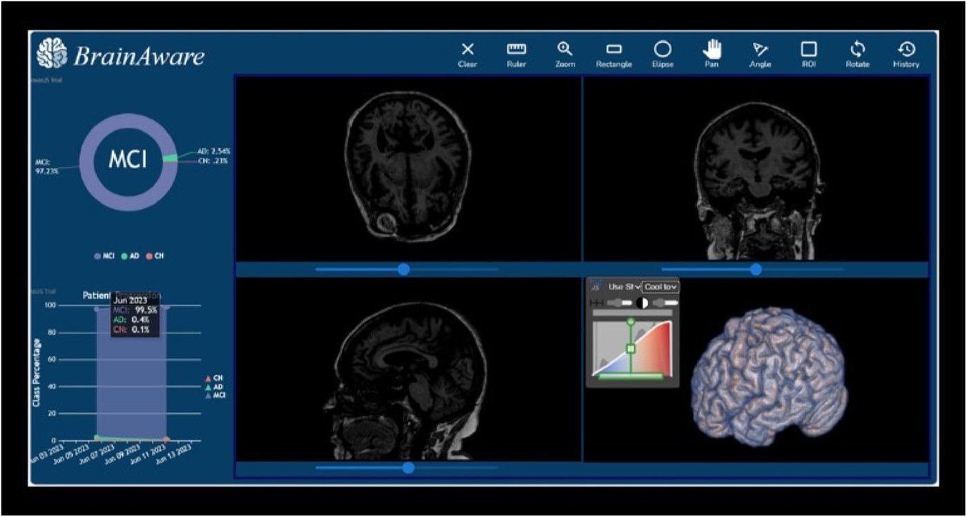
BrainAware takes a T1-weighted brain MRI, extracts the axial slices that contain the hippocampus, runs each slice through a fine-tuned VGG16 and averages the class probabilities. This page shows the viewer and the preprocessing on a few open-data scans.
Prediction
Live predictions coming soon.
Model input
150×150, as fed to the model (same orientation as training)
What the model sees
Each axial slice in the hippocampus range is skull stripped, cropped to the brain and resized to 150×150. The model classifies each slice on its own and averages the probabilities. Use the slider or the axial view to step through the slices.
About this demo
- Scans are open IXI T1 MRI of healthy volunteers, not ADNI.
- Preprocessing is approximated without FSL: affine registration to MNI152 (SimpleITK), MNI brain mask for skull stripping, Harvard-Oxford atlas for the hippocampus.
- Static page: no uploads, no server, no tracking. The full app needs the Django backend.
- Research only, not for clinical use.
How it works
- Data. ADNI T1-weighted MRI, 1,598 subjects (339 AD, 452 CN, 807 MCI).
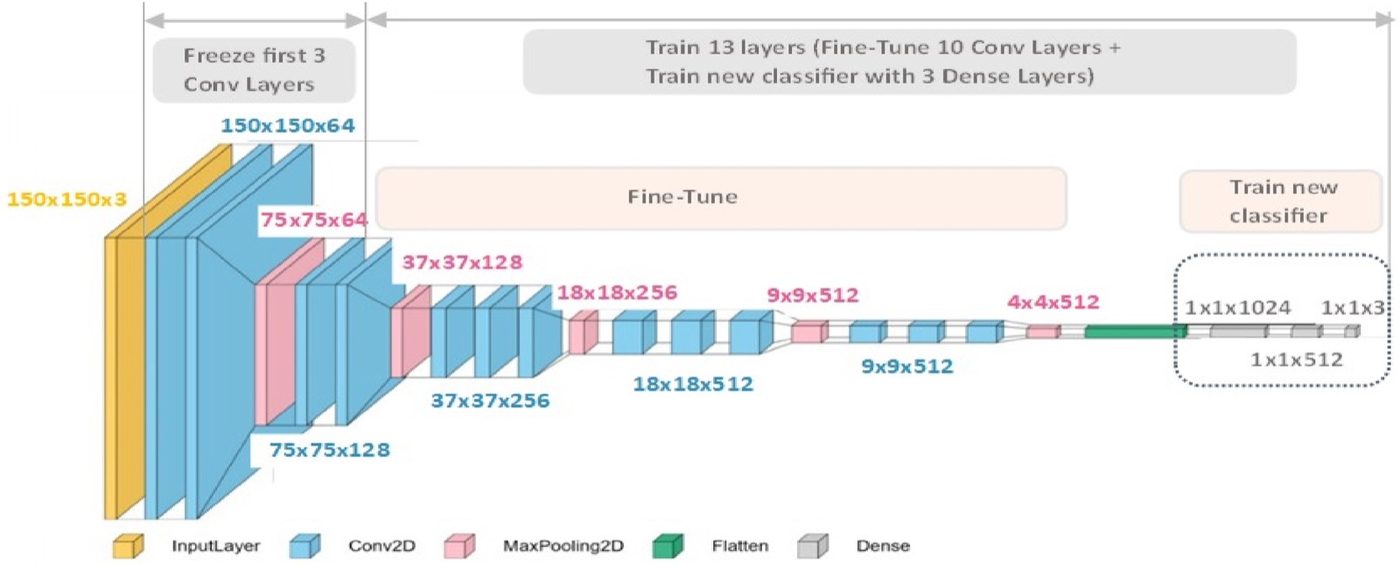
- Preprocessing. Register to MNI152, segment the hippocampus, skull strip, remove background, keep the slices that contain the hippocampus, crop to the brain, resize to 150×150.
- Model. VGG16 pretrained on ImageNet. First 3 conv layers frozen, the rest fine-tuned, new head with Dense 1024 → 512 → 3 (softmax).
- Training. Adam, exponential LR decay (0.96), class weights for the imbalance, progressive augmentation (strength increased in steps during training).
- Inference. Predict each slice, average the probabilities over slices, report CN / MCI / AD percentages.
Paper results (ADNI)
| Split | Accuracy | Loss |
|---|---|---|
| Train | 99.62% | 0.019 |
| Validation | 97.52% | 0.086 |
| Test (10-fold CV) | 98.17% | 0.077 |
| Class | Precision | Recall | F1 |
|---|---|---|---|
| AD | 0.99 | 0.96 | 0.98 |
| CN | 0.98 | 0.99 | 0.99 |
| MCI | 0.97 | 0.99 | 0.98 |
These numbers are slice-level results on ADNI. Reported in the paper on the ADNI test set; they are not results on the scans shown on this page. External validation is still open work.
Team
Built by Mohamed Hafez, Noha Tarek, Caroline Magdy, Mohammed Kamel and Alaa Melek. Supervised by Ibrahim Sadek. Co-authors: Lamees N. Mahmoud and Shafiq Ul Rehman.
Mohammed Abdelhalim on the paper is Mohamed Hafez.
Citation
Rehman S.U., Tarek N., Magdy C., Kamel M., Abdelhalim M., Melek A., Mahmoud L.N., Sadek I. AI-based tool for early detection of Alzheimer's disease. Heliyon 10(8), e29375, 2024. doi:10.1016/j.heliyon.2024.e29375
@article{rehman2024alzheimer,
  title   = {AI-based tool for early detection of Alzheimer's disease},
  author  = {Rehman, Shafiq Ul and Tarek, Noha and Magdy, Caroline and Kamel, Mohammed and
             Abdelhalim, Mohammed and Melek, Alaa and Mahmoud, Lamees N. and Sadek, Ibrahim},
  journal = {Heliyon},
  volume  = {10},
  number  = {8},
  pages   = {e29375},
  year    = {2024},
  doi     = {10.1016/j.heliyon.2024.e29375}
}
Data and credits
- MRI: IXI dataset (Imperial College London, Guy's, Hammersmith, Institute of Psychiatry), licensed CC BY-SA 3.0. The volumes and meshes on this page are derived from IXI T1 scans (bias corrected, registered, resampled) and are shared under the same license.
- Template: MNI ICBM152 2009a nonlinear symmetric (McConnell Brain Imaging Centre). Hippocampus mask: Harvard-Oxford subcortical atlas (via nilearn).
- 3D rendering: three.js (MIT).
- No ADNI data is published here. The model was trained on ADNI; access to ADNI is through adni.loni.usc.edu.